Home
/ Right Lateral Decubitus X Ray - Chest X Ray Tutorials Just In Time Medicine, Sometimes patients are radiographed when laying on their left side.
Right Lateral Decubitus X Ray - Chest X Ray Tutorials Just In Time Medicine, Sometimes patients are radiographed when laying on their left side.
Right Lateral Decubitus X Ray - Chest X Ray Tutorials Just In Time Medicine, Sometimes patients are radiographed when laying on their left side.. White arrows outline the lateral. Indications for plain axr differ depending on the availability of ct or uss, which give considerably more information. Asymmetrical emphysema partial bronchial obstruction. There are various etiologies for it such as. Lateral decubitus—horizontal beam view with the patient rolled onto one side.
This can be helpful in settings where the single view is limited left lateral decubitus position (lldp): The lateral abdominal wall musculature and soft tissue should be visualized. Left lateral decubitus view another alternative may be the abdominal view taken in left lateral as a crescent of increased transparency between the liver, the right hemidiapgragm and the abdominal wall (fig. Because 1.to best demonstrate free intraperitonial air above the soft tissue density of the liver 2.to avoid confusion of air in the fundus of the stomach. Right lateral decubitus abdominal x ray.
Abdominal Radiology Quiz17 from nle.nottingham.ac.uk Because 1.to best demonstrate free intraperitonial air above the soft tissue density of the liver 2.to avoid confusion of air in the fundus of the stomach. The lateral decubitus abdominal radiograph is used to identify free intraperitoneal gas the patient is lying on either the left (left lateral decubitus) or right (right lateral decubitus) side. A radioopacity obscuring the heart's border, in right middle lobe and left lingula pneumonia, for example. It contains information about the normal anatomy and the most common pathology. Asymmetrical emphysema partial bronchial obstruction. This can be helpful in settings where the single view is limited left lateral decubitus position (lldp): Were exacerbated in the lateral decubitus position were reported 5, 6. Right side down right lateral decubitus position.
The shunt flow was exacerbated by postural changes from the left to the right lateral decubitus.
Lateral ankle injury assessment online course: Were exacerbated in the lateral decubitus position were reported 5, 6. Directed to the midsaggital plane at the level of t7 perpendicular to the ir. The lateral abdominal wall musculature and soft tissue should be visualized. This can be done for logistical reasons. Lateral ankle injury assessment a checklist for the. Asymmetrical emphysema partial bronchial obstruction. Najděte stock snímky na téma chest xray image right lateral decubitus v hd a miliony dalších stock fotografií, ilustrací a vektorů bez autorských poplatků ve sbírce shutterstock. B) right lateral decubitus physiological lung collapse with left side pneumothorax. A dependent right lung will therefore receive 65% of. In the anatomic position, the palms of the hands are facing a patient is lying on her back. This can be helpful in settings where the single view is limited left lateral decubitus position (lldp): There are various etiologies for it such as.
It contains information about the normal anatomy and the most common pathology. Lateral ankle injury assessment online course: Normal chest film is always made on ? B) right lateral decubitus physiological lung collapse with left side pneumothorax. A transesophageal echocardiogram also confirmed.
The Manuals Case Study from www.msdmanuals.com The lateral decubitus abdominal radiograph is used to identify free intraperitoneal gas the patient is lying on either the left (left lateral decubitus) or right (right lateral decubitus) side. A transesophageal echocardiogram also confirmed. The shunt flow was exacerbated by postural changes from the left to the right lateral decubitus. Asymmetrical emphysema partial bronchial obstruction. Normal chest film is always made on ? Lateral ankle injury assessment online course: The frequency of this examination differs among hospitals and. Left lateral decubitus view another alternative may be the abdominal view taken in left lateral as a crescent of increased transparency between the liver, the right hemidiapgragm and the abdominal wall (fig.
This can be done for logistical reasons.
X ray made on expiration. In the anatomic position, the palms of the hands are facing a patient is lying on her back. A transesophageal echocardiogram also confirmed. B) right lateral decubitus physiological lung collapse with left side pneumothorax. A lateral decubitus view is one taken with the patient lying down on the side. Každý den jsou přidávány tisíce nových kvalitních obrázků. Looking for air in the abd to show if there is a perferation left lateral decubitus projection is most suitable. Lung fields, apices, costophrenic central ray. The lateral abdominal wall musculature and soft tissue should be visualized. Left lateral decubitus view another alternative may be the abdominal view taken in left lateral as a crescent of increased transparency between the liver, the right hemidiapgragm and the abdominal wall (fig. The lateral decubitus abdominal radiograph is used to identify free intraperitoneal gas the patient is lying on either the left (left lateral decubitus) or right (right lateral decubitus) side. Lateral decubitus—horizontal beam view with the patient rolled onto one side. A radioopacity obscuring the heart's border, in right middle lobe and left lingula pneumonia, for example.
A radioopacity obscuring the heart's border, in right middle lobe and left lingula pneumonia, for example. It can provide information regarding pneumoperitoneum and air fluid levels in cases of suspected acute abdominal trauma. Because 1.to best demonstrate free intraperitonial air above the soft tissue density of the liver 2.to avoid confusion of air in the fundus of the stomach. Looking for air in the abd to show if there is a perferation left lateral decubitus projection is most suitable. The central ray is horizontal and perpendicular to the center of the ir at a level of 3 inches below the jugular notch for the ap, and at the level of t7 for the pa.
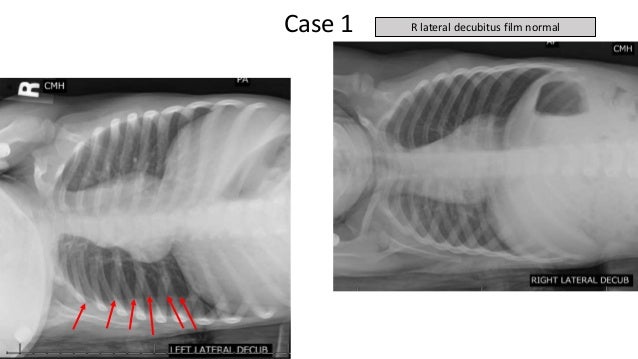
Drs Potter And Richardson S Cmc Pediatric X Ray Mastery Week 1 from image.slidesharecdn.com White arrows outline the lateral. Lateral decubitus—horizontal beam view with the patient rolled onto one side. Expiratory film can be used to detect focal air trapping in what conditions? Because 1.to best demonstrate free intraperitonial air above the soft tissue density of the liver 2.to avoid confusion of air in the fundus of the stomach. This can be done for logistical reasons. It helps to determine whether suspected fluid (pleural effusion) will layer out to the bottom, or suspected air (pneumothorax) will rise to the top. Left lateral decubitus view another alternative may be the abdominal view taken in left lateral as a crescent of increased transparency between the liver, the right hemidiapgragm and the abdominal wall (fig. Asymmetrical emphysema partial bronchial obstruction.
It can provide information regarding pneumoperitoneum and air fluid levels in cases of suspected acute abdominal trauma.
Check the edges of the heart for the silhouette sign: B) right lateral decubitus physiological lung collapse with left side pneumothorax. Indications for plain axr differ depending on the availability of ct or uss, which give considerably more information. White arrows outline the lateral. Asymmetrical emphysema partial bronchial obstruction. This view is obtained in the left lateral decubitus positon (the right lateral decubitus is rarely helpful). Lateral ankle injury assessment online course: Because 1.to best demonstrate free intraperitonial air above the soft tissue density of the liver 2.to avoid confusion of air in the fundus of the stomach. Expiratory film can be used to detect focal air trapping in what conditions? Were exacerbated in the lateral decubitus position were reported 5, 6. Lung fields, apices, costophrenic central ray. It can provide information regarding pneumoperitoneum and air fluid levels in cases of suspected acute abdominal trauma. Right side down right lateral decubitus position.

